Seeds of Science

154824818_22980b9cc5_oOCTOBER, 1889. Scientists flocked to Berlin for the annual meeting of the German Anatomical Society. The roster read like a who’s who of famous scientists of the day.

Into the fray marched a little-known Spaniard who’d spent years in Valencia and, later, Barcelona improving upon a method that made neurons visible under a microscope. Thanks to his patient tinkering, the Spaniard could see neurons in all their delicate, branching intricacy. He wanted to share his discoveries with other scientists. As he’d later say, he “gathered together for the purpose all my scanty savings and set out, full of hope, for the capital of the German Empire.”

In those days, scientific meetings were different from the parade of slideshows and posters sessions that they are today. The scientists at the 1889 meeting first read aloud from their papers and then took to their microscopes for demonstrations. The Spaniard unpacked his specimens and put them under several microscopes for the circulating scientists to view. Few came to see, in part because they expected little from a Spaniard. Spain was no scientific powerhouse. It lacked the scientific infrastructure and resources of countries like Germany, England, and France. What could one of its humble scientists possibly contribute to the meeting?

For the few curious gents who did stop by his demonstration, the Spaniard described his technique in broken French. Then he stepped aside and let them peer into the microscopes. Those who did became converts. The specimens spoke for themselves. Clear and complete, they revealed the intricate microarchitecture of neural structures like the retina and cerebellum.

Prominent German anatomists immediately adopted his technique and the Spaniard’s name quickly became known throughout the scientific community.

That name was Santiago Ramón y Cajal.Cajal

Ask any neuroscientist for his or her hero in the field and you are likely to hear this very name. Many consider him the founder of neurobiology as we know it today. The observations he made with his improved technique for seeing neurons allowed him to resolve a major controversy of the time and show that neurons are separate cells (as opposed to one huge, connected net). For his work, he won the Nobel Prize in Physiology or Medicine in 1906.

In short, he was an amazing guy who did amazing things – even though he wasn’t born in a wealthy nation known for science. Luckily, Cajal was able to get the tools and resources he needed to do his work. But what if he’d lived elsewhere, somewhere without the funds or equipment he needed? How far would that have set neuroscience back?

When I recently read an account of Cajal’s visit to Berlin, I found myself asking these questions. They reminded me of a Boston-based organization that is trying to equip the Cajals of today. The organization, a non-profit called Seeding Labs, partners with scientists, universities, and biomedical companies to equip stellar labs around the globe. (Full disclosure: The founder of Seeding Labs is the daughter of a family friend, which is how I first learned about the organization.)

The group’s core idea makes a lot of sense. Well-funded labs in the U.S. and other wealthy nations tend to update to newer models of their equipment often. These labs often discard perfectly functional older models that would be invaluable to scientists in developing nations. I’ve witnessed this kind of waste at major American universities. In the rush of doing science, people don’t have the time or energy to find new homes for their old autoclaves. They don’t even realize there’s a reason to try. While Seeding Labs now runs several programs to advance science in developing nations, its original aim was simply to turn one lab’s trash into another lab’s treasure.

I’m sure some struggling postdoc or assistant professor will read this post and scoff. Why devote energy to helping scientists in developing nations when we have a glut of scientists and a dearth of grants right here at home? It’s certainly true that research funding in America has tanked in recent years – a fact that needs to change. But in some countries the need is so great that a secondhand centrifuge could mean the difference between disappointment and discovery. That’s a pretty decent return on investment.

Here’s another benefit: labs in developing nations may be studying different problems than we are. They might focus on addressing local health or environmental concerns that we aren’t even aware of. So while scientists in wealthy nations find themselves racing to publish about well-trodden topics before competing labs, people in other countries may be researching crucial problems that wouldn’t otherwise be addressed.

And who knows? Perhaps these scientists are a good investment, in part, because of their relative isolation. Maybe a little distance from the scientific fray promotes ingenuity, creativity, and some good-old-fashioned tinkering. It certainly worked for Cajal.

____

Source: Stevens, Leonard A. Explorers of the Brain. Alfred A. Knopf, New York, 1971.

First photo credit: baigné par le soleil on Flickr, used via Creative Commons license

Second photo credit: Anonymous [Public domain], via Wikimedia Commons

Stuff and Brains Part 2: How Tools Come In Handy

298571748_c18ca5d78b_bHumans learn about objects by exploring them. I once described how my infant daughter explored objects, discovering their uses and properties through trial and error, observation, and plenty of dead ends. Her modest experiments illustrated a more universal truth: that from our earliest moments, our experience with objects in the world is fundamentally tied to our senses, to the ways we physically interact with them, and to the purposes they serve.

Last week I wrote about object-selective cortex, the part of visual cortex that lets us recognize people and stuff. I mentioned that this swath of the brain is speckled with several areas that specialize in processing certain object classes (e.g., faces, bodies, and scenes). If you consider object-selective cortex as a whole, you find that these specialized areas fit within a broader organization based on whether the to-be-recognized object is animate (a living, moving thing) or, if not, whether it’s large or small. While this may sound like a wacky way to divvy up object recognition, I mentioned some plausible reasons why your brain might map objects this way.

That’s the big-picture view. But what happens if we zoom in and explore one little bit of object-selective cortex in detail? Would we see a meaningful organization at this scale too? The answer, dear reader, is yes. In fact, this type of micro-organization can tell us volumes about how we recognize, understand, and use the objects around us.

For a beautiful example, let’s travel to the extrastriate body area (EBA).* The EBA is involved in visually recognizing bodies. Your EBA is active when you see a human body, regardless of whether the body is clothed or unclothed. It’s also active when you see parts of a body or even (to a lesser degree) when you see abstract body representations like stick figures. In 2010, scientists from Northumbria University used fMRI to ‘zoom in’ on the EBA in the left hemisphere. The team found that a chunk of the left EBA is specifically interested in pictures of hands, as opposed to other parts of the body. In essence, they found a micro-organization within the EBA, segregating hands from other body parts.

Before we talk more about hands, let’s visit another object-selective area in the same vicinity: the tool-selective area on the middle temporal gyrus. No kidding, your visual cortex has areas devoted to tools! The tool area on the middle temporal gyrus is engaged when you see a picture of a tool, be it a hammer, a stapler, or a fork. Patients with brain damage in this region tend to have trouble recalling information about the actions paired with common tools. But what counts as a tool for this region? One research group tried to answer this question by training adult subjects to use unfamiliar objects as tools. Using fMRI, the group showed that pictures of these objects activated the tool area after but not before training. In short, the brain dynamically reorganizes object recognition, or at least tool recognition, based on new experiences with objects.

But the story doesn’t end there. In 2012, the same group that discovered the hand area reported another find: that the hand area and the tool area overlap – a lot. What does this overlap mean? In essence, the same spot of cortex is active both when you see a hand and when you see a screwdriver or a pair of scissors. Notice that this goes against the broad divisions mentioned in my last post, since hands are animate and screwdrivers are not. Here, scale makes all the difference. When you zoom out, you see that object-selective cortex is broadly divvied up based on object animacy and size, but these divisions aren’t absolute and ubiquitous. Up close, you can find tiny bits of cortex that buck the trend, each with its own idiosyncratic combination of preferences.

Screen Shot 2013-10-24 at 3.39.27 PM

Figure from Bracci et al, 2012, showing the overlap of hand and tool areas in the left hemispheres of all but one of their subjects. Each slice represents the overlap (shown in cyan) in a different subject.

While each local mix of preferences may be idiosyncratic, it is probably not accidental. To save space and speed up reactions, brain organization is very well optimized. Chances are good that hands and tools overlap in the brain for a reason. But what might that reason be? It might stem from the fact that hands are intimately linked with tools in your visual experience. Since hands grip tools, you tend to see them together. You also tend to see faces and bodies together (that is, unless you’re watching a horror film.) And as it turns out, the face area and the body area on the bottom temporal surface of the right hemisphere appear to partially overlap as well. Could this be because faces and bodies, like hands and tools, tend to co-occur in our visual experience? It’s possible. Humans are quite sensitive to the statistical properties of our experience with objects.

But there’s another, quite different explanation for why faces overlap with bodies and tools overlap with hands in object-selective cortex. Brain organization tends to be dictated by where information needs to go next. (In essence, how the information will be used). The 2012 paper presents evidence that the overlapping hand/tool area is communicating with other areas of the brain that guide object-directed actions. The paper also cites another fMRI study that suggests the overlapping face and body areas in the right hemisphere communicate with parts of the brain involved in social interactions. In short, recognizing either a face or a body provides information that the social regions in your brain may need, while visual information about hands or tools may be invaluable when it comes time for reaching, grabbing, lifting, or stapling stuff.

Hands and tools. Faces and bodies. These are just a small sample of the many kinds of objects and creatures we see every day of our lives. Just imagine if we knew the micro-organization of every millimeter of object-selective cortex. Now that would be a map, one you started shaping from your earliest days on this earth. It would be a record of your lifetime of adventures with people and with stuff.

______

*Is it just me or does this post seem like an episode of The Magic School Bus?

Photo credits

Hands photo: Carmen Maria on Flickr

Brain images: Bracci et al, 2012 in The Journal of Neurophysiology

Bracci S, Cavina-Pratesi C, Ietswaart M, Caramazza A, & Peelen MV (2012). Closely overlapping responses to tools and hands in left lateral occipitotemporal cortex. Journal of neurophysiology, 107 (5), 1443-56 PMID: 22131379

Mapping a World of Stuff onto the Brain

4872199920_660cd8fb05_bWhile we have five wonderful senses, humans rely most on our sense of sight. The allocation of real estate in the brain reflects this hegemony; a far greater chunk of your cerebral cortex is dedicated to vision than to any other sense. So when you encounter people, objects, and animals in the world, you typically use visual information to tell your lover from a toothbrush from your cat. And while it would be reasonable to expect your brain to process all of these items in the same way, it does nothing of the sort. Instead, the visual cortex segregates and plays favorites.

The most dramatic examples of this segregation occur whenever you look at other people.  Within the large chunk of  visual cortex dedicated to object recognition, two areas in each hemisphere specifically process faces (the FFA and OFA) and two areas in each hemisphere specifically process bodies (the FBA and EBA). In each case, one of these areas is located on the side of the brain (near the back) while the other is tucked away on the bottom surface of the temporal lobe. It’s clear that these areas are important for recognizing faces and bodies. Damage to the face area FFA can profoundly impair one’s ability to recognize faces, while direct electrical stimulation of the same area can temporarily distort perception of a face. And when scientists used a magnetic pulse to momentarily disrupt activity in either the face area OFA or the body area EBA of healthy adults, their participants had difficulty discriminating between similar faces or similar bodies, respectively.

Yet the segregation of objects in your visual cortex doesn’t end there. Scientists have long known that visual information about scenes – including the landmarks and buildings that often define them – is processed separately as well. In fact we have at least two scene areas per hemisphere in classic visual cortex: one on the side of the brain (TOS) and one on the lower surface (PPA).*

But what about other types of objects? If you looked at pictures of a trampoline, a screwdriver, a lamppost, and a toad, would they follow the same path through your visual cortex? The answer is no. In a recent study, Talia Konkle and Alfonso Caramazza at Harvard showed people pictures of a wide range of animals and objects while scanning them with fMRI. They studied the activations in visual cortex for each image and used them to compute something they called preference maps. The preference maps indicated whether each bit of cortex preferred animals or objects and, separately, small or large things. When they combined these maps they found zones of visual cortex that preferred large objects, small objects, or animals of any size.** For large objects and animals (with two zones each), one zone was located on the side of the brain and the other on the lower surface. The only zone that preferred small objects over both large ones and animals lay right at the edge of the brain, smack dab between the side of the brain and its lower surface. The face and body areas fit almost entirely within the animal zones, while the scene areas lay within the large-object zones.

Screen Shot 2013-10-17 at 11.43.06 AM

Figure from Konkle & Caramazza, 2013 showing where the face areas, body areas, scene areas, and ‘preference zones’ were in one participant. Each gray blob represents the right hemisphere of the brain, with the left side of each blob representing the back of the brain. The top two brains show a side view while the bottom two show the bottom surface of the cortex.

It may seem odd that object representation in visual cortex is organized based on such arbitrary dimensions. Why should it matter whether the thing you see is big or small, made of cotton or has a cottontail? The study’s authors argue that these divisions make sense if one considers the various ways we use different types of objects. For instance, small objects are generally useful because you can manipulate and interact with them. Recognizing an apple, axe, or comb allows you to eat, chop, or fix your ‘do, respectively – so long as the visual information about these objects gets passed along to brain areas involved in reaching and grasping movements.

Objects like buildings, trees, or couches are obviously too large to be lifted or manipulated. Since they stay put, you’re more likely to use them as landmarks to help you navigate through a neighborhood, park, or room. But you can only use these objects this way if you send the visual information about them to brain regions involved in navigation.

Finally, we have living things, which can move, bite, and behave unpredictably. While a large animal like an elephant might trample you, a small one like a venomous spider or snake could be more lethal still. And don’t even get me started on people! In short, an animal’s size doesn’t determine how you will or won’t interact with it; you need to be ready to predict any animal’s behavior and react accordingly. Should you pet that dog or run from it? Communication between the animal-preferring zones of visual cortex and the social prediction centers in your brain might help you reach the right answer before it’s too late.

What’s the upshot of all this using, manipulating, predicting and fleeing? A wonderful and miraculous map of all the stuff in your world. It’s a modest little map – no larger than a napkin and half the thickness of an iPhone 5 – that represents a vast array of creatures, things, and people based on what they mean to you. How frickin’ amazing is that?

* I’ll get back to this mysterious pattern in a future post.

** I find it interesting that people generally approach the game Twenty Questions with the same category distinctions. The first two questions are almost invariably: Is it alive? And is it bigger than a breadbox?

___

Photo credits

Elephant and bird: Ludovic Hirlimann on Flickr, used via Creative Commons license

Figure with brains: Talia Konkle & Alfonso Caramazza in The Journal of Neuroscience

Konkle T, & Caramazza A (2013). Tripartite organization of the ventral stream by animacy and object size. The Journal of neuroscience : the official journal of the Society for Neuroscience, 33 (25), 10235-42 PMID: 23785139

Konkle T, & Caramazza A (2013). Tripartite organization of the ventral stream by animacy and object size The Journal of Neuroscience DOI: 10.1523/JNEUROSCI.0983-13.2013

Looking Schizophrenia in the Eye

272994276_3c83654e97_b
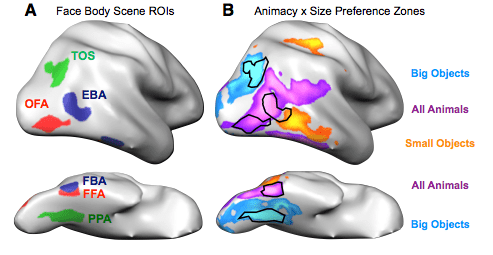
More than a century ago, scientists discovered something usual about how people with schizophrenia move their eyes. The men, psychologist and inventor Raymond Dodge and psychiatrist Allen Diefendorf, were trying out one of Dodge’s inventions: an early incarnation of the modern eye tracker. When they used it on psychiatric patients, they found that most of their subjects with schizophrenia had a funny way of following a moving object with their eyes.

When a healthy person watches a smoothly moving object (say, an airplane crossing the sky), she tracks the plane with a smooth, continuous eye movement to match its displacement. This action is called smooth pursuit. But smooth pursuit isn’t smooth for most patients with schizophrenia. Their eyes often fall behind and they make a series of quick, tiny jerks to catch up or even dart ahead of their target. For the better part of a century, this movement pattern would remain a mystery. But in recent decades, scientific discoveries have led to a better understanding of smooth pursuit eye movements – both in health and in disease.

Scientists now know that smooth pursuit involves a lot more than simply moving your eyes. To illustrate, let’s say a sexy jogger catches your eye on the street. When you first see the runner, your eyes are stationary and his or her image is moving across your retinas at some relatively constant rate. Your visual system (in particular, your visual motion-processing area MT) must first determine this rate. Then your eyes can move to catch up with the target and match its speed. If you do this well, the jogger’s image will no longer be moving relative to your retinas. From your visual system’s perspective, the jogger is running in place and his or her surroundings are moving instead. From both visual cues and signals about your eye movements, your brain can predict where the jogger is headed and keep moving your eyes at just the right speed to keep pace.

Although the smooth pursuit abnormalities in schizophrenia may sound like a movement problem, they appear to reflect a problem with perception. Sensitive visual tests show that motion perception is disrupted in many patients. They can’t tell the difference between the speeds of two objects or integrate complex motion information as well as healthy controls. A functional MRI study helped explain why. The study found that people with schizophrenia activated their motion-processing area MT less than controls while doing motion-processing tasks. The next logical question – why MT doesn’t work as well for patients – remains unanswered for now.

In my last two posts I wrote about how delusions can develop in healthy people who don’t suffer from psychosis. The same is true of not-so-smooth pursuit. In particular, healthy relatives of patients with schizophrenia tend to have jerkier pursuit movements than subjects without a family history of the illness. They are also impaired at some of the same motion-processing tests that stymie patients. This pattern, along with the results of twin studies, suggests that smooth pursuit dysfunction is inherited. Following up on this idea, two studies have compared subjects’ genotypes with the inheritance patterns of smooth pursuit problems within families. While they couldn’t identify exactly which gene was involved (a limitation of the technique), they both tracked the culprit gene to the same genetic neighborhood on the sixth chromosome.

http://www.youtube.com/watch?v=gqCgzSSwPLk

Despite this progress, the tale of smooth pursuit in schizophrenia is more complex than it appears. For one, there’s evidence that smooth pursuit problems differ for patients with different forms of the disorder. Patients with negative symptoms (like social withdrawal or no outward signs of emotion) may have problems with the first step of smooth pursuit: judging the target’s speed and moving their eyes to catch up. Meanwhile, those with more positive symptoms (like delusions or hallucinations) may have more trouble with the second step: predicting the future movement of the target and keeping pace with their eyes.

It’s also unclear exactly how common these problems are among patients; depending on the study, as many as 95% or as few as 12% of patients may have disrupted smooth pursuit. The studies that found the highest rates of smooth pursuit dysfunction in patients also found rates as high as 19% for the problems among healthy controls. These differences may boil down to the details of how the eye movements were measured in the different experiments. Still, the studies all agreed that people with schizophrenia are far more likely to have smooth pursuit problems than healthy controls. What the studies don’t agree on is how specific these problems are to schizophrenia compared with other psychiatric illnesses. Some studies have found smooth pursuit abnormalities in patients with bipolar disorder and major depression as well as in their close relatives; other studies have not.

Despite these messy issues, a group of scientists at the University of Aberdeen in Scotland recently tried to tell whether subjects had schizophrenia based on their eye movements alone. In addition to smooth pursuit, they used two other measures: the subject’s ability to fix her gaze on a stable target and how she looked at pictures of complex scenes. Most patients have trouble holding their eyes still in the presence of distractors and, when shown a meaningful picture, they tend to look at fewer objects or features in the scene.

Taking the results from all three measures into account, the group could distinguish between a new set of patients with schizophrenia and new healthy controls with an accuracy of 87.8%. While this rate is high, keep in mind that the scientists removed real-world messiness by selecting controls without other psychiatric illnesses or close relatives with psychosis. This makes their demonstration a lot less impressive – and a lot less useful in the real world. I don’t think this method will ever become a viable alternative to diagnosing schizophrenia based on their clinical symptoms, but the approach may hold promise in a similar vein: identifying young people who are at risk for developing the illness. Finding these individuals and helping them sooner could truly mean the difference between life and death.

_____

Photo credit: Travis Nep Smith on Flickr, used via Creative Commons License

Benson PJ, Beedie SA, Shephard E, Giegling I, Rujescu D, & St Clair D (2012). Simple viewing tests can detect eye movement abnormalities that distinguish schizophrenia cases from controls with exceptional accuracy. Biological psychiatry, 72 (9), 716-24 PMID: 22621999

Delusions: Making Sense of Mistaken Senses

6738201_646b9e485b_o

For a common affliction that strikes people of every culture and walk of life, schizophrenia has remained something of an enigma. Scientists talk about dopamine and glutamate, nicotinic receptors and hippocampal atrophy, but they’ve made little progress in explaining psychosis as it unfolds on the level of thoughts, beliefs, and experiences. Approximately one percent of the world’s population suffers from schizophrenia. Add to that the comparable numbers of people who suffer from affective psychoses (certain types of bipolar disorder and depression) or psychosis from neurodegenerative disorders like Alzheimer’s disease. All told, upwards of 3% of the population have known psychosis first-hand. These individuals have experienced how it transformed their sensations, emotions, and beliefs. Why hasn’t science made more progress explaining this level of the illness? What have those slouches at the National Institute of Mental Health been up to?

There are several reasons why psychosis has proved a tough nut to crack. First and foremost, neuroscience is still struggling to understand the biology of complex phenomena like thoughts and memories in the healthy brain. Add to that the incredible diversity of psychosis: how one psychotic patient might be silent and unresponsive while another is excitable and talking up a storm. Finally, a host of confounding factors plague most studies of psychosis. Let’s say a scientist discovers that a particular brain area tends to be smaller in patients with schizophrenia than healthy controls. The difference might have played a role in causing the illness in these patients, it might be a direct result of the illness, or it might be the result of anti-psychotic medications, chronic stress, substance abuse, poor nutrition, or other factors that disproportionately affect patients.

So what’s a well-meaning neuroscientist to do? One intriguing approach is to study psychosis in healthy people. They don’t have the litany of confounding experiences and exposures that make patients such problematic subjects. Yet at first glance, the approach seems to have a fatal flaw. How can you study psychosis in people who don’t have it? It sounds as crazy as studying malaria in someone who’s never had the bug.

In fact, this approach is possible because schizophrenia is a very different illness from malaria or HIV. Unlike communicable diseases, it is a developmental illness triggered by both genetic and environmental factors. These factors affect us all to varying degrees and cause all of us – clinically psychotic or not – to land somewhere on a spectrum of psychotic traits. Just as people who don’t suffer from anxiety disorders can still differ in their tendency to be anxious, nonpsychotic individuals can differ in their tendency to develop delusions or have perceptual disturbances. One review estimates that 1 to 3% of nonpsychotic people harbor major delusional beliefs, while another 5 to 6% have less severe delusions. An additional 10 to 15% of the general population may experience milder delusional thoughts on a regular basis.

Delusions are a common symptom of schizophrenia and were once thought to reflect the poor reasoning abilities of a broken brain. More recently, a growing number of physicians and scientists have opted for a different explanation. According to this model, patients first experience the surprising and mysterious perceptual disturbances that result from their illness. These could be full-blown hallucinations or they could be subtler abnormalities, like the inability to ignore a persistent noise. Patients then adopt delusions in a natural (if misguided) attempt to explain their odd experiences.

An intriguing study from the early 1960s illustrates how rapidly delusions can develop in healthy subjects when expectations and perceptions inexplicably conflict. The study, run on twenty college students at the University of Copenhagen, involved a version of the trick now known as the rubber hand illusion. Each subject was instructed to trace a straight line while his or her hand was inside a box with a secret mirror. For several trials, the subject watched his or her own hand trace the line correctly. Then the experimenters surreptitiously changed the mirror position so that the subject was now watching someone else’s hand trace the straight line – until the sham hand unexpectedly veered off to the right! All of the subjects experienced the visible (sham) hand as their own and felt that an involuntary movement had sent it off course. After several trials with this misbehaving hand, the subjects offered explanations for the deviation. Some chalked it up to their own fatigue or inattention while others came up with wilder, tech-based explanations:

 . . . five subjects described that they felt something strange and queer outside themselves, which pressed their hand to the right or resisted their free mobility. They suggested that ‘magnets’, ‘unidentified forces’, ‘invisible traces under the paper’, or the like, could be the cause.

In other words, delusions may be a normal reaction to the unexpected and inexplicable. Under strange enough circumstances, anyone might develop them – but some of us are more likely to than others.

My next post will describe a clever experiment that planted a delusion-like belief in the heads of healthy subjects and used trickery and fMRI to see how it influenced some more than others. So stay tuned. In the meantime, you may want to ask yourself which members of your family and friends are prone to delusional thinking. Or ask yourself honestly: could it be you?

_______

Photo credit: MiniTar on Flickr, available through Creative Commons