Outsourcing Memory

3088541520_00a0721cde_b

Do you rely on your spouse to remember special events and travel plans? Your coworker to remember how to submit some frustrating form? Your cell phone to store every phone number you’ll ever need? Yeah, me too. You might call this time saving or delegating, but if you were a fancy psychologist you’d call it transactive memory.

Transactive memory is a wonderful concept. There’s too much information in this world to know and remember. Why not store some of it in “the cloud” that is your partner or coworker’s brain or in “the cloud” itself, whatever and wherever that is? The idea of transactive memory came from the innovative psychologist Daniel Wegner, most recently of Harvard, who passed away in July of this year. Wegner proposed the idea in the mid-80s and framed it in terms of the “intimate dyad” – spouses or other close couples who know each other very well over a long period of time.

Transactive memory between partners can be a straightforward case of cognitive outsourcing. I remember monthly expenses and you remember family birthdays. But it can also be a subtler and more interactive process. For example, one spouse remembers why you chose to honeymoon at Waikiki and the other remembers which hotel you stayed in. If the partners try to recall their honeymoon together, they can produce a far richer description of the experience than if they were to try separately.

Here’s an example from a recent conversation with my husband. It began when my husband mentioned that a Red Sox player once asked me out.

“Never happened,” I told him. And it hadn’t. But he insisted.

“You know, years ago. You went out on a date or something?”

“Nope.” But clearly he was thinking of something specific.

I thought really hard until a shred of a recollection came to me. “I’ve never met a Red Sox player, but I once met a guy who was called up from the farm team.”

My husband nodded. “That guy.”

But what interaction did we have? I met the guy nine years ago, not long before I met my husband. What were the circumstances? Finally, I began to remember. It wasn’t actually a date. We’d gone bowling with mutual friends and formed teams. The guy – a pitcher – was intensely competitive and I was the worst bowler there. He was annoyed that I was ruining our team score and I was annoyed that he was taking it all so seriously. I’d even come away from the experience with a lesson: never play games with competitive athletes.

Apparently, I’d told the anecdote to my husband after we met and he remembered a nugget of the story. Even though all of the key details from that night were buried somewhere in my brain, I’m quite sure that I would never have remembered them again if not for my husband’s prompts. This is a facet of transactive memory, one that Wegner called interactive cueing.

In a sense, transactive memory is a major benefit of having long-term relationships. Sharing memory, whether with a partner, parent, or friend, allows you to index or back up some of that memory. This fact also underscores just how much you lose when a loved one passes away. When you lose a spouse, a parent, a sibling, you are also losing part of yourself and the shared memory you have with that person. After I lost my father, I noticed this strange additional loss. I caught myself wondering when I’d stopped writing stories on his old typewriter. I realized I’d forgotten parts of the fanciful stories he used to tell me on long drives. I wished I could ask him to fill in the blanks, but of course it was too late.

Memories can be shared with people, but they can also be shared with things. If you write in a diary, you are storing details about current experiences that you can access later in life. No spouse required. You also upload memories and information to your technological gadgets. If you store phone numbers in your cell phone and use bookmarks and autocomplete tools in your browser, you are engaging in transactive memory. You are able to do more while remembering less. It’s efficient, convenient, and downright necessary in today’s world of proliferating numbers, websites, and passwords.

In 2011, a Science paper described how people create transactive memory with online search engines. The study, authored by Betsy Sparrow, Jenny Liu, and Wegner, received plenty of attention at the time, including here and here.

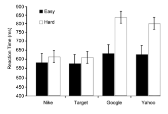
In one experiment, they asked participants either hard or easy questions and then had them do a modified Stroop task that involved reporting the physical color of a written word rather than naming the word. This was a measure of priming, essentially whether a participant has been thinking about that word or similar concepts recently. Sometimes the participants were tested with the names of online search engines (Google, Yahoo) and at others they were tested with other name brands (Nike, Target). After hard questions, the participants took much longer to do the Stroop task with Google and Yahoo than with the other brand names, suggesting that hard questions made them automatically think about searching the Internet for the answer.

Screen Shot 2013-11-21 at 1.53.54 PM

The other experiments described in the paper showed that people are less likely to remember trivia if they believe they will be able to look it up later. When participants thought that items of trivia were saved somewhere on a computer, they were also more likely to remember where the items were saved than they were to remember the actual trivia items themselves. Together, the study’s findings suggest that people actively outsource memory to their computers and to the Internet. This will come as no surprise to those of us who can’t remember a single phone number offhand, don’t know how to get around without the GPS, and hop on our smartphones to answer the simplest of questions.

Search engines, computer atlases, and online databases are remarkable things. In a sense, we’d be crazy not to make use of them. But here’s the rub: the Internet is jam-packed with misinformation or near-miss information. Anti-vaxxers, creationists, global warming deniers: you can find them all on the web. And when people want the definitive answer, they almost always find themselves at Wikipedia. While Wikipedia has valuable information, it is not written and curated by experts. It is not always the God’s-honest-truth and it is not a safe replacement for learning and knowing information ourselves. Of course, the memories of our loved ones aren’t foolproof either, but at least they don’t carry the aura of authority that comes with a list of citations.

Speaking of which. There is now a Wikipedia page for “The Google Effect” that is based on the 2011 Science article. A banner across the top shows an open book featuring a large question mark and the following warning: “This article relies largely or entirely upon a single source. . . . Please help improve this article by introducing citations to additional sources.” The citation for the first section is a dead link. The last section has two placeholders for citations, but in lieu of numbers they say, According to whom?

Folks, if that ain’t a reminder to be wary of the outsourcing your brain to Google and Wikipedia, I don’t know what is.

_________

Photo credits:

1. Photo by Mike Baird on Flickr, used via Creative Commons license

2. Figure from “Google Effects on Memory: Cognitive Consequences of Having Information at Our Fingertips” by Betsy Sparrow, Jenny Liu, and Daniel M. Wegner.

Sparrow B, Liu J, & Wegner DM (2011). Google effects on memory: cognitive consequences of having information at our fingertips. Science (New York, N.Y.), 333 (6043), 776-8 PMID: 21764755

Seeds of Science

154824818_22980b9cc5_oOCTOBER, 1889. Scientists flocked to Berlin for the annual meeting of the German Anatomical Society. The roster read like a who’s who of famous scientists of the day.

Into the fray marched a little-known Spaniard who’d spent years in Valencia and, later, Barcelona improving upon a method that made neurons visible under a microscope. Thanks to his patient tinkering, the Spaniard could see neurons in all their delicate, branching intricacy. He wanted to share his discoveries with other scientists. As he’d later say, he “gathered together for the purpose all my scanty savings and set out, full of hope, for the capital of the German Empire.”

In those days, scientific meetings were different from the parade of slideshows and posters sessions that they are today. The scientists at the 1889 meeting first read aloud from their papers and then took to their microscopes for demonstrations. The Spaniard unpacked his specimens and put them under several microscopes for the circulating scientists to view. Few came to see, in part because they expected little from a Spaniard. Spain was no scientific powerhouse. It lacked the scientific infrastructure and resources of countries like Germany, England, and France. What could one of its humble scientists possibly contribute to the meeting?

For the few curious gents who did stop by his demonstration, the Spaniard described his technique in broken French. Then he stepped aside and let them peer into the microscopes. Those who did became converts. The specimens spoke for themselves. Clear and complete, they revealed the intricate microarchitecture of neural structures like the retina and cerebellum.

Prominent German anatomists immediately adopted his technique and the Spaniard’s name quickly became known throughout the scientific community.

That name was Santiago Ramón y Cajal.Cajal

Ask any neuroscientist for his or her hero in the field and you are likely to hear this very name. Many consider him the founder of neurobiology as we know it today. The observations he made with his improved technique for seeing neurons allowed him to resolve a major controversy of the time and show that neurons are separate cells (as opposed to one huge, connected net). For his work, he won the Nobel Prize in Physiology or Medicine in 1906.

In short, he was an amazing guy who did amazing things – even though he wasn’t born in a wealthy nation known for science. Luckily, Cajal was able to get the tools and resources he needed to do his work. But what if he’d lived elsewhere, somewhere without the funds or equipment he needed? How far would that have set neuroscience back?

When I recently read an account of Cajal’s visit to Berlin, I found myself asking these questions. They reminded me of a Boston-based organization that is trying to equip the Cajals of today. The organization, a non-profit called Seeding Labs, partners with scientists, universities, and biomedical companies to equip stellar labs around the globe. (Full disclosure: The founder of Seeding Labs is the daughter of a family friend, which is how I first learned about the organization.)

The group’s core idea makes a lot of sense. Well-funded labs in the U.S. and other wealthy nations tend to update to newer models of their equipment often. These labs often discard perfectly functional older models that would be invaluable to scientists in developing nations. I’ve witnessed this kind of waste at major American universities. In the rush of doing science, people don’t have the time or energy to find new homes for their old autoclaves. They don’t even realize there’s a reason to try. While Seeding Labs now runs several programs to advance science in developing nations, its original aim was simply to turn one lab’s trash into another lab’s treasure.

I’m sure some struggling postdoc or assistant professor will read this post and scoff. Why devote energy to helping scientists in developing nations when we have a glut of scientists and a dearth of grants right here at home? It’s certainly true that research funding in America has tanked in recent years – a fact that needs to change. But in some countries the need is so great that a secondhand centrifuge could mean the difference between disappointment and discovery. That’s a pretty decent return on investment.

Here’s another benefit: labs in developing nations may be studying different problems than we are. They might focus on addressing local health or environmental concerns that we aren’t even aware of. So while scientists in wealthy nations find themselves racing to publish about well-trodden topics before competing labs, people in other countries may be researching crucial problems that wouldn’t otherwise be addressed.

And who knows? Perhaps these scientists are a good investment, in part, because of their relative isolation. Maybe a little distance from the scientific fray promotes ingenuity, creativity, and some good-old-fashioned tinkering. It certainly worked for Cajal.

____

Source: Stevens, Leonard A. Explorers of the Brain. Alfred A. Knopf, New York, 1971.

First photo credit: baigné par le soleil on Flickr, used via Creative Commons license

Second photo credit: Anonymous [Public domain], via Wikimedia Commons

Another Time, Another Place

7052169001_4ac18f1ef6_b

Whenever I visit my childhood home outside of Chicago I try to make it to the local pancake house. The buttery pancakes would be reason enough, but they’re not the only reason I stop by. A stroll through that pancake house is truly a stroll down memory lane. Each table I pass triggers a memory of a meal shared with different people in different decades of my life. One moment I’m eating German pancakes with my college boyfriend. The next, I am passing menus to my new husband’s family.  The next, I am celebrating my eighth grade graduation with my parents and older brother.

Memories return you to a specific time and place. Consider so-called flashbulb memories, or vivid memories of  dramatic moments that caught you off-guard. I remember exactly where I was when I heard that a plane had struck one of the Twin Towers and, later, when I learned that my father had died. I remember that I was sitting on the living room rug in my Somerville apartment when I watched Columbia transform from a space shuttle into a streak of fire across the sky. Is it helpful to remember where I was sitting? Not in the slightest. But in the murky, mysterious realm of memory, when and what are inextricably linked with where.

Mention the word “memory” to neuroscientists and you’re sure to get them thinking of the hippocampus, a sliver of tissue nestled deep inside each hemisphere of the brain. The hippocampus has been synonymous with memory since the late 1950s, when William Scoville and Brenda Milner described a patient who was incapable of forming new memories after both of his hippocampi were removed. Since then, throngs of neuroscientists have devoted their careers to studying the hippocampus. Among other revelations, they’ve discovered a class of neurons called place cells that represent (you guessed it) information about place.

How do cells represent place? To illustrate, let’s say you’re in your favorite coffee shop. Some of the place cells in your hippocampus will fire like crazy when you walk through the entrance. Others save their enthusiasm until you are waiting in line to order your latte, stopping at the counter for milk and sugar, or settling in at your favorite table. When you physically occupy their place-of-interest, they go nuts – like a neural alarm signaling your location. At this moment, you are here!

The same principle applies to my experience at the pancake house. Different place cells fire at different tables. In essence, these sets of cells provide a unique neural code for each space I can occupy in the restaurant. And this code has been with me for a while. When I sat in the corner booth after my graduation from middle school, I formed a memory of that celebration that included the code for that particular spot. Decades later, sitting in that booth or even walking past it can trigger a similar code in my brain, one that elicits the rest of that dusty old memory.

While eternally cool, place cells have become old news in hippocampal research. The new hippocampal hotness is studying “time cells”. These recently discovered neurons prefer to fire at different intervals after an event (say, ten seconds versus one minute after you step into the coffee shop). This research fad is a bit amusing, as it turns out that place cells and “time cells” are one and the same. This fact hasn’t stopped scientists from referring to “time cells,” but it has forced them to typically use the term in quotation marks.

As scientists studied the time code in the hippocampal cells of rats, a flaw in their experiments became clear. Their studies recorded the neural activity of moving rats, which means that the firing patterns observed by the scientists could reflect changes in time, changes in the rat’s location, or in its motion.

Two recent papers addressed this issue and clarified the nature of “time cells” in the hippocampus. The first of these appeared in the journal Neuron in June of this year. The paper, by Benjamin Kraus, Michael Hasselmo, and collaborators at Boston University, describes an experiment that has as much to do with your time spent sweating it out at the gym as it does with your memory of past events. The scientists recorded the activity of hippocampal cells in rats as they ran on a treadmill or moved around in a simple maze. Since the rat remained in the same location as it ran on the treadmill, the researchers could decouple the rat’s location from the passage of time and the distance the rat ran. Since the authors could vary the speed of the treadmill, they could also piece apart the related variables of time and distance.

The scientists found that “time cells” still produced a time code when location was kept constant (on the treadmill). Using some fancy modeling, they also showed that the activity of most “time cells” reflected a combination of elapsed time and distance run, but a smaller number of “time cells” seemed to care only about time or distance. They also found that these same cells behaved like normal place cells when the rat walked around a simple maze. In short, place cells (a.k.a. “time cells”) can convey information about place, time, and distance travelled to varying degrees that also change under different conditions.

A second paper on the subject came out in a September issue of The Journal of Neuroscience. The authors, Christopher MacDonald, Howard Eichenbaum*, and colleagues (also from Boston University) eliminated the variable of location by physically restraining the rats from moving with a special headpiece that attached to the rats’ heads. This headpiece locked into the testing apparatus so that the rats couldn’t move their heads during testing. Unlike the fitness buff rats in the prior study, these rats were given a memory task. They got a whiff of an odor and then another whiff of an odor a few seconds later. If the second odor was the same as the first, the rat licked its waterspout and got a reward (a drop of water). If the two odors were different, the rat was not supposed to lick.

Even though the rats were completely immobile, the rats’ “time cells” showed a strong time code. Different cells fired at different times during the delay. These cells also seemed to represent what information (in this case, the odors presented for the task). The scientists found that the overall pattern of “time cell” firing was more similar when the rats remembered the same odor than when they remembered different odors across trials.

In short, place/time cells can represent what, when, and where in a variety of ways, depending on a variety of factors. This representation is flexible – just as memory must be in order for you to remember the date of your anniversary, the feel of your first kiss, and the items on your next shopping list. The remarkable thing about memory is that it is both flexible and robust, meaning that it is resistant to degradation or being swamped out by noise. It can return us to times, places, and experiences that are far away and decades past. For that, we can thank the hippocampus, neural codes, and a set of remarkable cells with an identity crisis.

_____

Photo credit: Stu Rapley on Flickr, used via Creative Commons License

*Howard Eichenbaum was also a middle author on the Neuron paper. Much of the recent work on “time cells” has come from his lab and affiliated labs at Boston University.

Kraus BJ, Robinson RJ 2nd, White JA, Eichenbaum H, & Hasselmo ME (2013). Hippocampal “time cells”: time versus path integration. Neuron, 78 (6), 1090-1101 PMID: 23707613

MacDonald CJ, Carrow S, Place R, & Eichenbaum H (2013). Distinct hippocampal time cell sequences represent odor memories in immobilized rats. The Journal of Neuroscience : the official journal of the Society for Neuroscience, 33 (36), 14607-14616 PMID: 24005311

Stuff and Brains Part 2: How Tools Come In Handy

298571748_c18ca5d78b_bHumans learn about objects by exploring them. I once described how my infant daughter explored objects, discovering their uses and properties through trial and error, observation, and plenty of dead ends. Her modest experiments illustrated a more universal truth: that from our earliest moments, our experience with objects in the world is fundamentally tied to our senses, to the ways we physically interact with them, and to the purposes they serve.

Last week I wrote about object-selective cortex, the part of visual cortex that lets us recognize people and stuff. I mentioned that this swath of the brain is speckled with several areas that specialize in processing certain object classes (e.g., faces, bodies, and scenes). If you consider object-selective cortex as a whole, you find that these specialized areas fit within a broader organization based on whether the to-be-recognized object is animate (a living, moving thing) or, if not, whether it’s large or small. While this may sound like a wacky way to divvy up object recognition, I mentioned some plausible reasons why your brain might map objects this way.

That’s the big-picture view. But what happens if we zoom in and explore one little bit of object-selective cortex in detail? Would we see a meaningful organization at this scale too? The answer, dear reader, is yes. In fact, this type of micro-organization can tell us volumes about how we recognize, understand, and use the objects around us.

For a beautiful example, let’s travel to the extrastriate body area (EBA).* The EBA is involved in visually recognizing bodies. Your EBA is active when you see a human body, regardless of whether the body is clothed or unclothed. It’s also active when you see parts of a body or even (to a lesser degree) when you see abstract body representations like stick figures. In 2010, scientists from Northumbria University used fMRI to ‘zoom in’ on the EBA in the left hemisphere. The team found that a chunk of the left EBA is specifically interested in pictures of hands, as opposed to other parts of the body. In essence, they found a micro-organization within the EBA, segregating hands from other body parts.

Before we talk more about hands, let’s visit another object-selective area in the same vicinity: the tool-selective area on the middle temporal gyrus. No kidding, your visual cortex has areas devoted to tools! The tool area on the middle temporal gyrus is engaged when you see a picture of a tool, be it a hammer, a stapler, or a fork. Patients with brain damage in this region tend to have trouble recalling information about the actions paired with common tools. But what counts as a tool for this region? One research group tried to answer this question by training adult subjects to use unfamiliar objects as tools. Using fMRI, the group showed that pictures of these objects activated the tool area after but not before training. In short, the brain dynamically reorganizes object recognition, or at least tool recognition, based on new experiences with objects.

But the story doesn’t end there. In 2012, the same group that discovered the hand area reported another find: that the hand area and the tool area overlap – a lot. What does this overlap mean? In essence, the same spot of cortex is active both when you see a hand and when you see a screwdriver or a pair of scissors. Notice that this goes against the broad divisions mentioned in my last post, since hands are animate and screwdrivers are not. Here, scale makes all the difference. When you zoom out, you see that object-selective cortex is broadly divvied up based on object animacy and size, but these divisions aren’t absolute and ubiquitous. Up close, you can find tiny bits of cortex that buck the trend, each with its own idiosyncratic combination of preferences.

Screen Shot 2013-10-24 at 3.39.27 PM

Figure from Bracci et al, 2012, showing the overlap of hand and tool areas in the left hemispheres of all but one of their subjects. Each slice represents the overlap (shown in cyan) in a different subject.

While each local mix of preferences may be idiosyncratic, it is probably not accidental. To save space and speed up reactions, brain organization is very well optimized. Chances are good that hands and tools overlap in the brain for a reason. But what might that reason be? It might stem from the fact that hands are intimately linked with tools in your visual experience. Since hands grip tools, you tend to see them together. You also tend to see faces and bodies together (that is, unless you’re watching a horror film.) And as it turns out, the face area and the body area on the bottom temporal surface of the right hemisphere appear to partially overlap as well. Could this be because faces and bodies, like hands and tools, tend to co-occur in our visual experience? It’s possible. Humans are quite sensitive to the statistical properties of our experience with objects.

But there’s another, quite different explanation for why faces overlap with bodies and tools overlap with hands in object-selective cortex. Brain organization tends to be dictated by where information needs to go next. (In essence, how the information will be used). The 2012 paper presents evidence that the overlapping hand/tool area is communicating with other areas of the brain that guide object-directed actions. The paper also cites another fMRI study that suggests the overlapping face and body areas in the right hemisphere communicate with parts of the brain involved in social interactions. In short, recognizing either a face or a body provides information that the social regions in your brain may need, while visual information about hands or tools may be invaluable when it comes time for reaching, grabbing, lifting, or stapling stuff.

Hands and tools. Faces and bodies. These are just a small sample of the many kinds of objects and creatures we see every day of our lives. Just imagine if we knew the micro-organization of every millimeter of object-selective cortex. Now that would be a map, one you started shaping from your earliest days on this earth. It would be a record of your lifetime of adventures with people and with stuff.

______

*Is it just me or does this post seem like an episode of The Magic School Bus?

Photo credits

Hands photo: Carmen Maria on Flickr

Brain images: Bracci et al, 2012 in The Journal of Neurophysiology

Bracci S, Cavina-Pratesi C, Ietswaart M, Caramazza A, & Peelen MV (2012). Closely overlapping responses to tools and hands in left lateral occipitotemporal cortex. Journal of neurophysiology, 107 (5), 1443-56 PMID: 22131379

Mapping a World of Stuff onto the Brain

4872199920_660cd8fb05_b
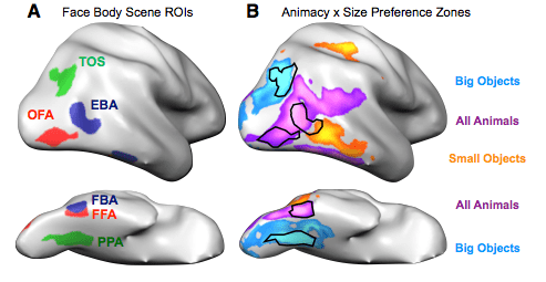
While we have five wonderful senses, humans rely most on our sense of sight. The allocation of real estate in the brain reflects this hegemony; a far greater chunk of your cerebral cortex is dedicated to vision than to any other sense. So when you encounter people, objects, and animals in the world, you typically use visual information to tell your lover from a toothbrush from your cat. And while it would be reasonable to expect your brain to process all of these items in the same way, it does nothing of the sort. Instead, the visual cortex segregates and plays favorites.

The most dramatic examples of this segregation occur whenever you look at other people.  Within the large chunk of  visual cortex dedicated to object recognition, two areas in each hemisphere specifically process faces (the FFA and OFA) and two areas in each hemisphere specifically process bodies (the FBA and EBA). In each case, one of these areas is located on the side of the brain (near the back) while the other is tucked away on the bottom surface of the temporal lobe. It’s clear that these areas are important for recognizing faces and bodies. Damage to the face area FFA can profoundly impair one’s ability to recognize faces, while direct electrical stimulation of the same area can temporarily distort perception of a face. And when scientists used a magnetic pulse to momentarily disrupt activity in either the face area OFA or the body area EBA of healthy adults, their participants had difficulty discriminating between similar faces or similar bodies, respectively.

Yet the segregation of objects in your visual cortex doesn’t end there. Scientists have long known that visual information about scenes – including the landmarks and buildings that often define them – is processed separately as well. In fact we have at least two scene areas per hemisphere in classic visual cortex: one on the side of the brain (TOS) and one on the lower surface (PPA).*

But what about other types of objects? If you looked at pictures of a trampoline, a screwdriver, a lamppost, and a toad, would they follow the same path through your visual cortex? The answer is no. In a recent study, Talia Konkle and Alfonso Caramazza at Harvard showed people pictures of a wide range of animals and objects while scanning them with fMRI. They studied the activations in visual cortex for each image and used them to compute something they called preference maps. The preference maps indicated whether each bit of cortex preferred animals or objects and, separately, small or large things. When they combined these maps they found zones of visual cortex that preferred large objects, small objects, or animals of any size.** For large objects and animals (with two zones each), one zone was located on the side of the brain and the other on the lower surface. The only zone that preferred small objects over both large ones and animals lay right at the edge of the brain, smack dab between the side of the brain and its lower surface. The face and body areas fit almost entirely within the animal zones, while the scene areas lay within the large-object zones.

Screen Shot 2013-10-17 at 11.43.06 AM

Figure from Konkle & Caramazza, 2013 showing where the face areas, body areas, scene areas, and ‘preference zones’ were in one participant. Each gray blob represents the right hemisphere of the brain, with the left side of each blob representing the back of the brain. The top two brains show a side view while the bottom two show the bottom surface of the cortex.

It may seem odd that object representation in visual cortex is organized based on such arbitrary dimensions. Why should it matter whether the thing you see is big or small, made of cotton or has a cottontail? The study’s authors argue that these divisions make sense if one considers the various ways we use different types of objects. For instance, small objects are generally useful because you can manipulate and interact with them. Recognizing an apple, axe, or comb allows you to eat, chop, or fix your ‘do, respectively – so long as the visual information about these objects gets passed along to brain areas involved in reaching and grasping movements.

Objects like buildings, trees, or couches are obviously too large to be lifted or manipulated. Since they stay put, you’re more likely to use them as landmarks to help you navigate through a neighborhood, park, or room. But you can only use these objects this way if you send the visual information about them to brain regions involved in navigation.

Finally, we have living things, which can move, bite, and behave unpredictably. While a large animal like an elephant might trample you, a small one like a venomous spider or snake could be more lethal still. And don’t even get me started on people! In short, an animal’s size doesn’t determine how you will or won’t interact with it; you need to be ready to predict any animal’s behavior and react accordingly. Should you pet that dog or run from it? Communication between the animal-preferring zones of visual cortex and the social prediction centers in your brain might help you reach the right answer before it’s too late.

What’s the upshot of all this using, manipulating, predicting and fleeing? A wonderful and miraculous map of all the stuff in your world. It’s a modest little map – no larger than a napkin and half the thickness of an iPhone 5 – that represents a vast array of creatures, things, and people based on what they mean to you. How frickin’ amazing is that?

* I’ll get back to this mysterious pattern in a future post.

** I find it interesting that people generally approach the game Twenty Questions with the same category distinctions. The first two questions are almost invariably: Is it alive? And is it bigger than a breadbox?

___

Photo credits

Elephant and bird: Ludovic Hirlimann on Flickr, used via Creative Commons license

Figure with brains: Talia Konkle & Alfonso Caramazza in The Journal of Neuroscience

Konkle T, & Caramazza A (2013). Tripartite organization of the ventral stream by animacy and object size. The Journal of neuroscience : the official journal of the Society for Neuroscience, 33 (25), 10235-42 PMID: 23785139

Konkle T, & Caramazza A (2013). Tripartite organization of the ventral stream by animacy and object size The Journal of Neuroscience DOI: 10.1523/JNEUROSCI.0983-13.2013